−Table of Contents
3D Tissue Organization Toolbox
Authors
We provide a toolbox to segment and analyze tissues in 3D.
Tissue Analysis Plugin is a toolbox which allow to analyze cells and
nuclei and explore the spatial structure of tissue.
This toolbox is developed by Thomas's group.The goal is to
suggest a workflow in order to choose a set of optimal methodologies
for quantification and modeling of tissue organization based on
3D microscopy images.
This ImageJ plugin toolbox is easy access to biologist. The tissue
toolbox was used to analyze endocrine cells in a paper:
“A novel toolbox to investigate tissue spatial organization
applied to the study of the islets of Langerhans”.
Scientific Report
TRAN THI NHU Hoa, ARROJO E DRIGO Rafael, BERGGREN Per-Olof, and
BOUDIER Thomas
Scientific Report
Image and Pervasive Access Lab
Email: hoa.tran_thi_nhu@etu.upmc.fr or Thomas Boudier
Introduction
Data Requirement
Tissue Analysis Toolbox requires some 3D microscopy images as input data:
* 3D Nuclei DAPI staining: nuclear labelling DAPI which capture nucleus in 3D.
* Markerset : a markerset represents a set of fluorescence markers or imaging channels used in an experiment. More than one markerset may be associated with a single tissue. This allows users to perform analysis with different combinations of the available markers. In our paper, we applied our toolbox with endocrine cells which composed of three channels (three markers): named insulin staining, glucagon staining, and somatostatin staining.
* Image file format:
- Only 8-bit or 16-bit TIFF format file of 3D are currently supported. All images in one dataset should have the same size XYZ.
- Input: A composite image composed of all different channels and one nuclei marker image.
* We also provide an example of input images extracted from islet of Langerhans: (more detail see reference paper)
- DAPI stainning nuclei image: C4-dapi.tif
- Three channels correspond to cytoplasmic (label or marker) and efficient to determine cell types: Somatostatin: C1-delta.tif, Insulin: C2-beta.tif, Glucagon: C3-alpha.tif
- To open these images example: launch ImageJ, open .zip file and obtain the .tiff format of these images.
Installation
Tissue Analysis Toolbox.
* Install ImageJ environment, see intruction in ImageJ web page. ImageJ version should be >= 1.50a
and Java 1.7 or later is required JDK
* Download the Tissue Analysis plugin in this link: tissueanalysis_.jar.
* To work with 3D TIFF image, some libraries are required also: imagescience.jar, imageware.jar.
* The ConvexHull3D plugin is required only for measurements: quickhull3d.jar.
* Java3D is required, download our 3D Viewer version 3D Viewer plugin
(version 4 or later) or go to the main website:3D Viewer
* Mcib3d core and plugin, version 3.8 or latter: mcib3d-core_3.83.jar, mcib3d_plugins_3.83.jar included many functions for 3D image processing.
* Package Lipid Droplet Counter
which contains 3D band pass filter, included in 3D Nuclei Segmentation of tissue organization plugin.
* Put the jar file into the plugin folder of ImageJ and refresh menu.
* More details about the required plugin you can find out here.
Starting Tissue Framework
* Launch ImageJ program.
* ImageJ –> Plugin –> 3D Tissue Spatial Analysis.
The source code of this framework you can find at our github repository:
https://github.com/nhuhoa/tissue_organization
Features
* Filtering
* Nuclei segmentation
* SLIC clustering
* Watershed segmentation
* Compute cell zone
* Compute cell type
* Analyze cellular interaction
* Spatial Statistic
* Clusters Analysis
* Visualization of graph

1. Pre-filtering and normalize labelling image.
Before processing the marker - labelling image, we need to filter and normalize intensity of image. ImageJ –> Plugin –> 3D Tissue Spatial Analysis–> Pre-filtering Input: a composite image which contain different channels and dapi nuclei image also. Input parameters: Choose the label correspond to each image. It can be M1, M2, M3 (markers, cytoplasmic to determine cell type) or DAPI (nuclei), or *NONE* if number of channels is smaller number of options provided. Output: filtered images of different channels and save it into folder. The output images will be save into a folder named “filtered” with the rename of images is: C1.tif, C2.tif, C3.tif, C4-dapi.tif.
Normalize image
Select image and input threshold value. The program will remove the voxel which have intensity value below this threshold and after the program will normalize image within the range [0, 255]. This step is enable to remove a part of noise in 3D microscopy image.

2. Nuclei Segmentation
The second step is to detect nuclei in the image by applying the algorithm of segmentation. Images corresponding to nuclear labelling (DAPI) were first processed by 3D median filter to reduce noise and homogenized intensities inside nuclei. After that image were then filtered by a 3D band pass filter which is the approximated diameter of the nuclei region.The output is a 32-bit image, we then scale it to a 16-bit image. Our plugin 3D Spot Segmentation was used to segment the nuclei, where local maxima from the band pass filtering are used for seeds.
A set of defaut parameters are provide in order to suggest the parameters for segmentation. But quality of nuclei image, volume of nucleus vary depend on different species and different tissue.
The toolbox enable you to adjust input parameters and observe the output.
ImageJ –> Plugin –> 3D Tissue Spatial Analysis –> NUCLEI SEGMENTATION
To adjust segmentation parameters:
Input image: DAPI nuclei image.
Pre-processing: choose pre-filtering (3D Median 4-4-2) if the image has not yet filtered.
Maximal Nucleus Size, Minimal Nucleus Size: you need to provide the range [min_size, max_size] of nucleus radius size in the image.
Seed Threshold: threshold using to detect seed in the Band Pass Filtered Image. Normally, we use the threshold of 30000 to extract the brightest voxel correspond to seed in the 16-bit image. If image is at low intensity, we can reduce this parameter.
For the detail of Spot Segmentation method, you can read the documentation of our previous work:
http://imagejdocu.tudor.lu/lib/exe/fetch.php?media=plugin:stacks:3d_ij_suite:3d_seg_spot_tutorial.pdf
Output: the output is a 16-bit image of nuclei segmentation which contain different regions. Each region is indexed by one value of intensity and have different color by applying LUT 3-3-2 RGB.
To observe the segmentation result:
+ Open segmented image in 3D Manager and 3D Viewer.
+ A window of 3D Manager will appear and show the list of objects, 3D Viewer show the visualization of objects in 3D.
+ From 3D Manager, you can check the 3D nucleus object profile and have manual validation: detect merge objects and split them.
See more of 3D Manager at the following link: http://imagejdocu.tudor.lu/doku.php?id=plugin:stacks:3d_roi_manager:start

3. Cell Zone Detection
The normal way to detect a cell is by using segmented nuclei as seeds inside membrane region.
But for 3D microscopy image, 3D membrane label is unavailable. In this work we apply wateshed - region growing technique to obtain the cell zone.
ImageJ –> Plugin –> 3D Tissue Spatial Analysis –> CELL ZONE
Input: nuclei segmented image.
Distance: count from border of segmented nucleus to outside. User can define this parameter based on observed cell.
If cells are very closes, boundary is counted haft way from maximum distance between border of two cells.
Output: watershed image, define cell zone around each nucleus as seed.

4. 3D SLIC Clustering
To combine information of different markers, we use 3D Multi-Voxels Simple Linear Iterative Clustering (SLIC) algorithm. This algorithm perform an unsupervised learning of different channels.
ImageJ –> Plugin –> 3D Tissue Spatial Analysis –> SLIC 3D CLUSTERING
Input: different channels - markerset. Choose number of input images.
Min size, Max size: [min_size, max_size] provide the range, which is the volume possible of each
clustering region. The defaut volume [100, 300] correspond to the max size and min size of nucleus in dataset, as volume of region that the program will use for clustering.
Nb iterations: number of iterations that program will take to perform clustering and refine the result of clustering. The defaut value is 10 iterations. The complexity of SLIC algorithm depend on the size XYZ of 3D image.
Output: one image which contain many regions, each region correspond to one cluster of voxels,
named label.tiff

5. 3D SLIC 3 Channels
We finally assign each zone to the channel having the highest median value inside the zone. If all the median values inside the zone are lower than predefined thresholds, the zone is assigned to the unlabelled type. An image is then created with the voxels having the value of their corresponding zone.
ImageJ –> Plugin –> 3D Tissue Spatial Analysis –> SLIC 3D 3 CHANNELS
Input: choose image correspond to different labels and SLIC label as result of SLIC 3D CLUSTERING.
Threshold : Input threshold value. If the median value of a SLIC region is below this value, this region can not take into account.
Output: SLIC with intensity value of region correspond to only one channel (one label) in the list of existing label (M1=1, M2=2, M3=3)

6. Cell Type Detection
ImageJ –> Plugin –> 3D Tissue Spatial Analysis –> CELL TYPE DETECTION
Method: take into account different region. Based on the intensity value of three channels and cell profile that we obtain from the previous step, we compute the cell type of each cell by quantify the marker inside each cell zone.
Input: a watershed - cell zone image, segmented nuclei image, and SLIC labelF image.
Region observed: INSIDE_OUTSIDE NUCLEUS, OUTSIDE NUCLEUS, INSIDE NUCLEUS, WATERSHED REGION
Input parameter: ratio of histogram of label
Min distance
Maximum distance inside of nuclei region take into account.
Maximum distance outside of nuclei region take into account.
Output: cell type image
Cell profile: Nuclei.zip and Region.zip
7. Cellular Interaction Computation
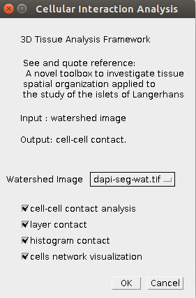
We define cell-cell contact based on nucleus zone. Two cells are neighbor if the nucleus zone of 2 cells are touching, means have the voxel contact. (user can handle the number of voxels contact as input parameter in the next version)
Input: watershed image, 3D ROI file Nuclei.zip, Regions.zip. At the current version our plugin will read 2 files: Nuclei.zip and Regions.zip are located in the same folder with watershed image.
Choose option: provide different options for computation: cell to cell contact, layer contact, histogram contact between beta cells and alpha cells, cells network visualization.
Output: excel files: AllContact.csv, LayerContact.csv, histogram_beta_alpha.csv
Image of cells network visualization.

8. Random Organization
The program will compare the cellular interaction between observed raw data and average of 100 simulated random organization model.
ImageJ –> Plugin –> 3D Tissue Spatial Analysis –> RANDOM ORGANIZATION
Input: watershed image.
Choose one of cell type observed: TYPE1, TYPE2, TYPE3
Choose source cell type observed: TYPE1, TYPE2, TYPE3
Number of random organization: by defaut is 100 times simulated random organization.
Output:
The number of interactions of observed raw data.
The number of interactions of 100 simulated models and average.
===== 9. Cluster Analysis =====
ImageJ –> Plugin –> 3D Tissue Spatial Analysis –> CLUSTER ANALYSIS
Input: watershed image.
Choose type cell observed: TYPE1, TYPE2, TYPE3
Choose whether to compute Clusters Spatial Statistic
Output: excel file Cluster.csv include the computation results:
index cluster, number of observed cells in each clusters, number of clusters, average cells in total clusters, max number of cells in the biggest cluster.
Graph of spatial statistic F-function and G-function of cluster and SDI value.

9. Spatial Statistic Analysis
Compute the spatial statistic of organization model.
ImageJ –> Plugin –> 3D Tissue Spatial Analysis –> SPATIAL STATISTIC
Input: watershed image, Nuclei.zip, Region.zip
Choose cell type observed: TYPE1, TYPE2, TYPE3
Choose cell type source: TYPE1, TYPE2, TYPE3
Choose type distance: Euclidean Distance, Cell Distance.
Choose model for compute spatial statistic: Raw Data, Random Organization, Clusters.
Choose spatial statistic function to compute: F-function, G-function.
See more about Spatial Statistic Plugin at the following link:
http://imagejdocu.tudor.lu/doku.php?id=plugin:analysis:spatial_statistics_2d_3d:start


