Table of Contents
MicroSCoBioJ
Introduction
MicroSCoBioJ is a collection of three plugins to create and visualize 3D fluorescence volume rendering
Authors
Fabio Frosi (frogsi (at) libero.it)
Giuseppe Vicidomini (gvicid (at) gwdg.de)
Features
Requires: ImageJ + Java 3D ( http://java3d.j3d.org/download.html )
Limitations: Only works with 8-bit images stack
Description
MicroSCoBioJ contains three plugins:
- Mesh Maker MicroSCoBioJ computes the triangles or tetrahedra mesh corresponding an user-defined treshold
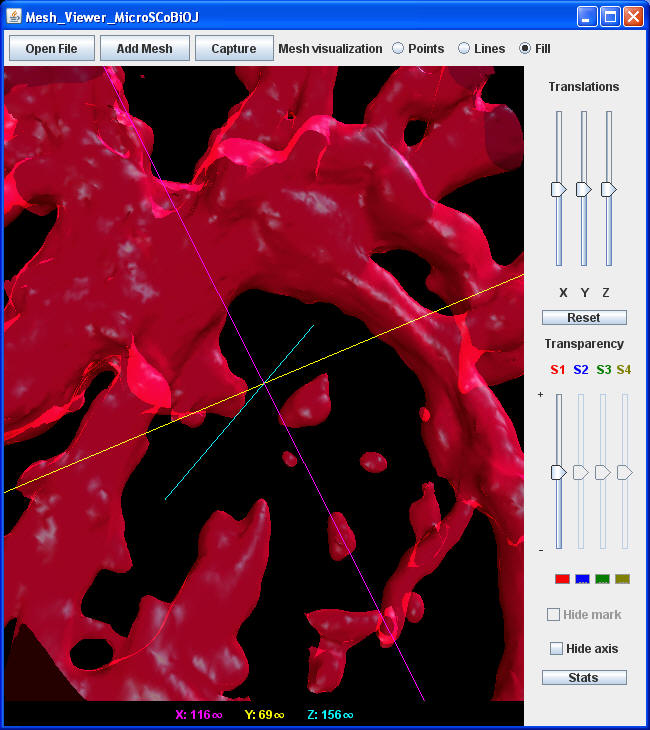
- Mesh Viewer MicroSCoBioJ 3D viewer of the mesh created by MeshMaker
- WAT MicroSCoBioJ Weight Adaptive Threshold
Mesh Maker MicroSCoBioJ
- Load a 8-bit images stack
- Start MeshMaker plugin ( Plugins >MicroSCoBioJ > MeshMaker MicroSCoBioJ )
- Using the following menu one can choose: treshold, voxel dimensions, and algorithm (marching cubes or marching tetratrahedra)
- The mesh calculation could take several minultes (up to an hour), a progress bar will display the percentage of the performed caculation.
- Save the mesh file
Mesh Viewer MicroSCoBioJ
- This plugin show up to four different meshes (with different colors: red, blue, green and yellow).
- It is possible to visualize the mesh as points, lines or fill surface.
- Other capabilities: translations, zoom, trasparency (indipendent for each mesh)
WAT MicroSCoBioJ
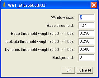
The algorithm used in the previous step, identifies a global threshold for all pixels of an image, using statistical properties of the image histogram. Unfortunately this algorithm is not able to accommodate changes in the distribution of different fluorescence concentration in a structure. Chow and Kaneko (ref. 1) solved this problem dynamically changing the threshold over the image: each local pixel threshold is obtained by statistical investigation of the intensity values of the local neighbourhoods of such pixel. We combined the IsoData threshold (Th_IsoD) (ref. 2), with the dynamic threshold (Thd), to find a weight adaptive threshold (Thwa): Thwa(x,y) = wIsoD* ThIsoD + wd *Thd(x,y) + wbase*Thbase+background with wIsoD + wd + wbase= 1 Thbase can be used to increase or decrease the final threshold on the base of prior information; moreover, further information, e.g. background, can be directly added to the algorithm.
Using WAT MicroScoBioJ menu one can choose the following parameters: the window size, Thbase , three weights and the background value.
References:
- Chow, C. K. & Kaneko, T. Automatic Boundary Detection of the Left Ventricle from Cineangiograms. Comp. Biomed. Res. 5, 338-410. (1972).
- Ridler, T. & Calvard, S. Picture thresholding using an iterative selection method. IEEE Trans. Systems Man Cybernet. 8, 629-632. (1978).
Installation
Download MicroSCoBiOJ.zip and unzip it into the plugins folder and restart ImageJ.
Download
microscobioj.zip 158KB


