−Table of Contents
Particle in Cell-3D
Particle in Cell-3D is an ImageJ Macro designed to quantify the uptake of macro- and nanoparticles by cells.
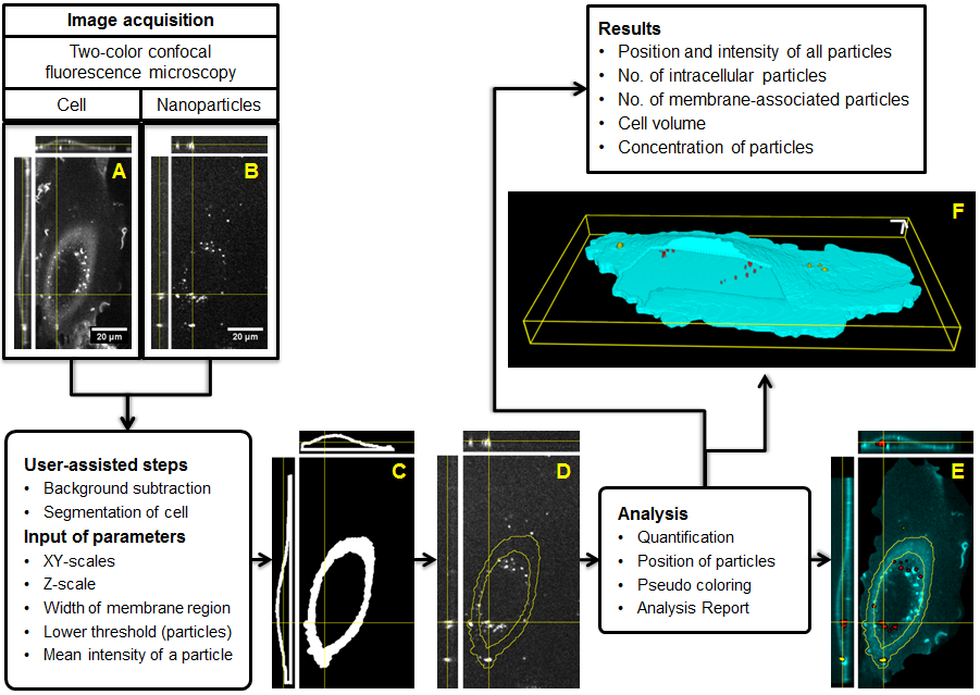
 Fig. 1: Particle_in_Cell-3D processing overview. a) Representative confocal section image and orthogonal projections of a HUVEC cell membrane stained with CellMask™ Deep Red. b) The respective image of silica nanoparticles labeled with perylene, a fluorescent dye. The 3D location of an intracellular particle is marked by the crossing yellow lines. c) A smoothing filter is applied and the image of the cell is transformed into a white mask. The image stack of masks is further processed to deliver a 3D reconstruction of the cell boundaries. Intracellular and membrane region are also defined in this step. d) The cell boundaries, or regions of interest, are then used to segment the image of the nanoparticles (yellow outline). The segmentation procedure occurs throughout the image stack, leading to a 3D localization of the particles with respect to the cell. e) Quantitative image analysis takes place. The intensity of each object (particle or agglomerate) is compared to the intensity of a single particle previously measured in a calibration procedure. Nanoparticles are pseudo-colored according to the cellular region. In this example the cell membrane is shown in cyan, the intracellular nanoparticles appear in red, and the membrane-associated nanoparticles in yellow. f) 3D representation of nanoparticle uptake after evaluation. Intracellular nanoparticles can be seen through the window intentionally open in the membrane region (cyan). 3D scale bars = 5 µm. (Taken from ref. [3].)
Input
Routines 1, 2 and 3:
- Confocal fluorescence image stack of a single cell with stained plasma membrane
- Respective image stack of nanoparticles
Routines 4 and 5:
- Confocal fluorescence image stack of nanoparticles
References
[1] Torrano A.A., Blechinger J., Osseforth C., Argyo C., Reller A., Bein T., Michaelis J., and Bräuchle C.
A fast analysis method to quantify nanoparticle uptake on a single cell level
Nanomedicine 8(11)(2013), 1815.[link]
[2] Blechinger J., Bauer A.T., Torrano A.A., Gorzelanny C., Bräuchle C., and Schneider S.W.
Uptake kinetics and nanotoxicity of silica nanoparticles are cell type dependent
Small 9(23)(2013), 3970.[link]
[3] Torrano A.A. & Bräuchle C.
Precise quantification of silica and ceria nanoparticle uptake revealed by 3D fluorescence microscopy
Beilstein Journal of Nanotechnology 5 (2014), 1616.[link]
Contact Information
Adriano A. Torrano
Department of Chemistry and Center for NanoScience (CeNS)
University of Munich (LMU), Germany
Adriano.Torrano'at'gmail.com
